Clinical reasoning: a logical process
Clinical reasoning: a logical process

A 70-year-old woman had a diagnosis of breast cancer several years earlier. In a recent visit the patient referred back pain and a difficulty to walk, without muscular strength in her legs.
Reasoning process
Representation heuristic
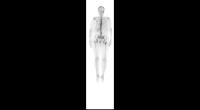
The medical oncologist that attended the patient at that moment, thought in a spinal cord compression as the first diagnostic possibility, a thought supported by the symptoms and the presence of metastases in the bone scintigraphy. The physical examination did not include a complete neurological assessment to determine if there was a sensitive level.
Subsequently, during the evolution, the initial diagnosis of spinal cord compression was not demonstrated.
Evolution
The patient was in a weak condition and she stayed in bed most of the time, although she could move the lower extremities at a thirty-degree angle and maintained strength, but could not stand without help. There was not a neurologic sensitive level.
The patient reported that she could not sit because the pain in her left hip, although an uptake increase was described on the right side of the bone scan.
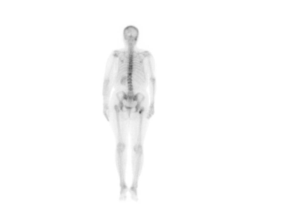
Right or left? The first thought of the doctor was to think that the report of the bone scan could be mistaken, because the pain was on the left side and the report informed that the metastases was on the right side. Or maybe, she thought, could it be a lytic lesion on the left side, usually not detected by the scan?
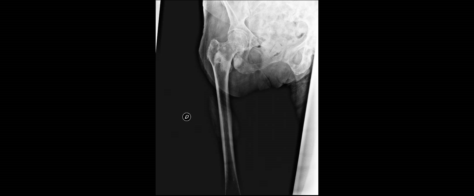
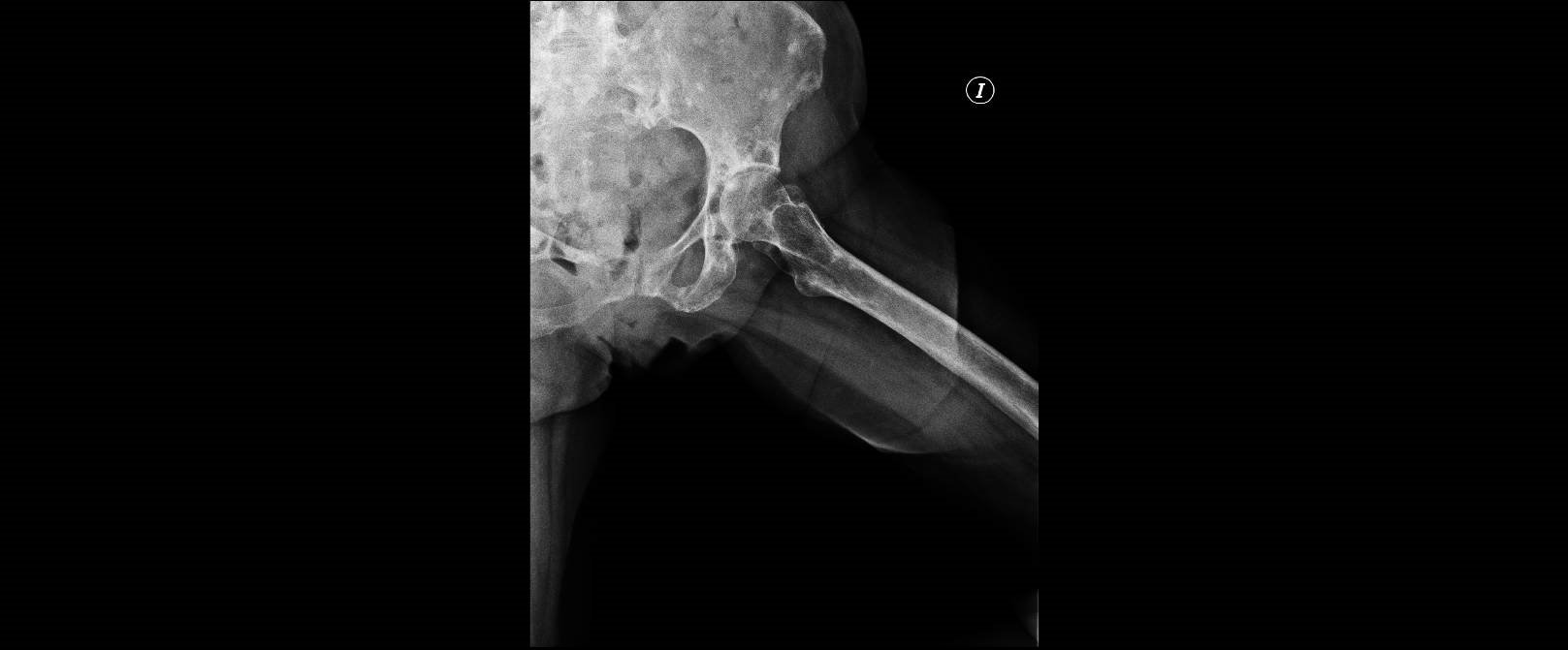
There was a mark to identify the side in the radiological image. After a consultation to the Nuclear Medicine Department it was confirmed that the lesion was located on the right side. An x-ray of both hips confirmed the presence of a lytic lesion in the left femur, and another blastic lesion in the right femur.
Commentary and Analysis
Some comments can help to increase the safety of this clinical scenario:
- It is important in a graphic medical report to clearly identify laterality. Normally until now it has been identified with a letter (R or L in English), but there is a change and the whole word, right or left, is now included. In the case of her bone scintigraphy, the image taken in “negative” may increase the possibility of an error.
2.Each diagnostic test has its specifications and sensitivity and specificity. In the case of bone scintigraphy, the absence of uptake in a pure lytic lesions is well known.
- Although there was no confirmation of the initial presumptive diagnosis , the doctor’s attitude was correct, since she applied a safety attitude considering the worst possible diagnostic scenario.
- When in doubt, the clinician should contact the doctor who performed the test.
- Always give priority to the patient’s symptoms, and assess the radiographic findings in this context.
Author: Lorenzo Alonso. Foro Osler/Prodiagnosis