Clinical reasoning and the Emperor new clothes: the problem with perception in Radiology

You look, but you don’t see
Vision is an automatic act, it keeps us aware of our environment to react before a danger or an opportunity. When we want to get more information, we concentrate our minds and our eyes over an object or situation, in a more conscious and active way. After analyzing an
object or a scene, we change our brain to the automatic visual mode again.There are then two actions: one, concentrating on a part of the visual field and the second one analysis and interpretation.
Nowadays, we have evolved in Radiology to the digital format, what is an advantage, but the size of the computer screen or the resolution have a direct influence on the perception. We could have a tendency to visualize only a limited area, giving origin to a perception bias, usually tunnel vision (a partial visualization of the visual field, usually in the center of the field) or satisfaction of search (stop looking for more details once you localize a finding that can explain some aspects of the problem).
Clinical case
A 52-year-old man with a syndrome of the obstructive biliary tract in association with a pancreatic cancer had a procedure to insert a catheter into his biliary tract. Six months later he started with nausea and vomiting because the lump was compressing the duodenum. His doctor decided to insert a prosthesis inside the duodenum to solve the obstruction.
When the doctor looked to the screen, she saw an intraabdominal catheter, in a correct central abdominal position, and she was happy about the situation.
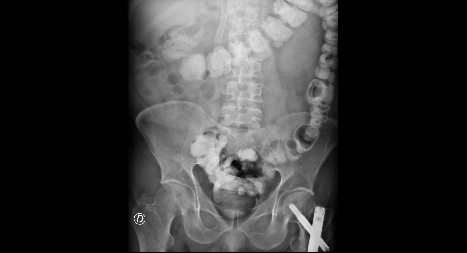
Practical exercise: look at the next picture during three seconds. Can you see the intraabdominal catheter? Close your eyes later, wait for five seconds and look back again, going up and down. Can you see something different?
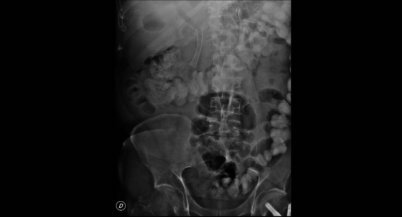
Now, look at the next picture. What do you think about the situation?
Final Comment
There were two catheters or prosthesis in the first picture, one in the upper abdominal area, what is the one located in the biliary tract. The second one is down, in the pelvis, after migrating from the duodenum . In the second picture you can see only the biliary catheter, because the prosthesis in the duodenum was expelled from the body.
The are two main perception biases in radiology:
- Tunnel vision: you just look for an area of the field and then you miss the information around.
- Satisfaction of search: when you find something, then you stop looking for more, because the image explain most of your reasoning.
Bibliography: you can see a free article about biases in radiology here