Radiology: different views of the same problem, but always in pain
Objective
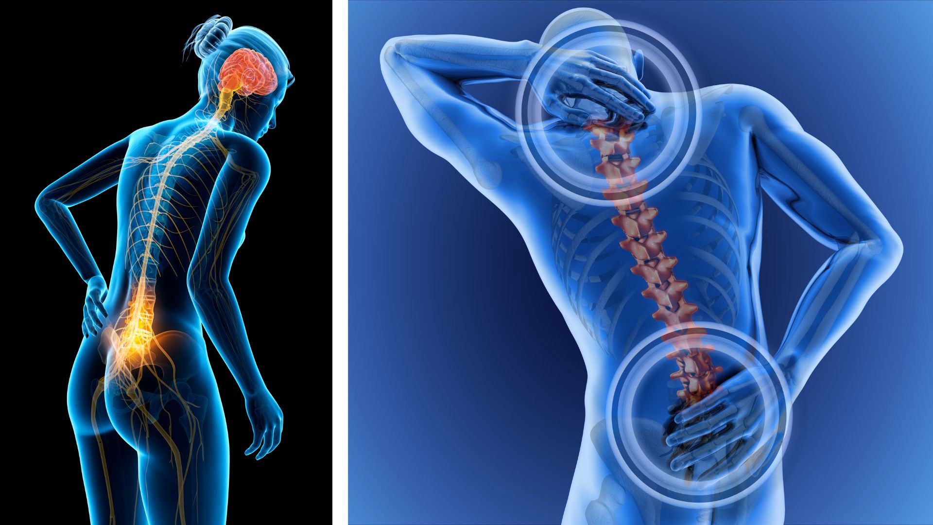
Bone metastases are common in several types of cancer, such as lung, breast, kidney or thyroid, with two main divisions between blastic or a lytic presentation. The knowledge of the presence of this situation is important to decide the best treatment for the patient, and to detect in advance the possibility of a bone fracture. A bone scan, a CT or a plain X-ray are useful tests to evidence if there is a metastasis, but pain is almost universally the main complaint of the patient.
Clinical case
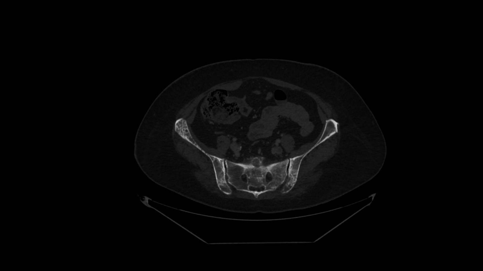
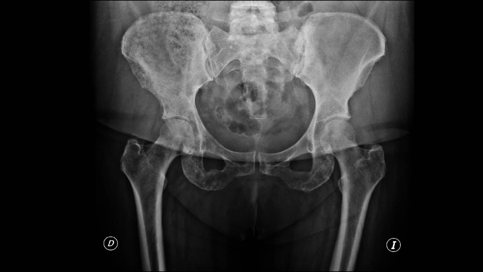
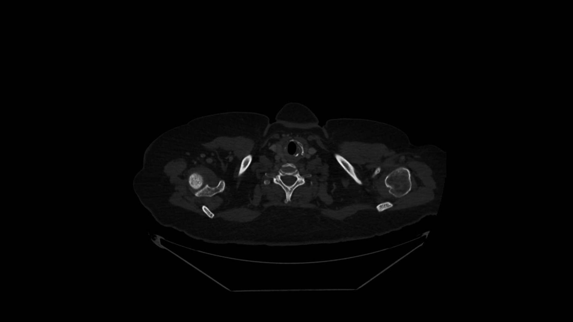
A fifty-two-year old woman with a previous diagnosis of breast cancer, started with pain in her right shoulder and in the right hip, always associated with the physical activity of the normal everyday life. We will show in the next images the radiological findings associated with two different radiological tools, and the conclusion that we can extract after a comparative analysis.

Comments
Bone metastases can be detected with a bone scan, a CT scan and a plain X-ray. A plain X-ray can give an important and additional information, for instance they can help to improve the idea about the situation of the long bones, in terms of coping with the weight of the patient or the physical activity of the everyday life. The conclusion is clear: a CT scan or a bone scan is a good test, but don´t forget to ask for a plain X-ray, mainly when the long bones are affected. And a very important clinical advice: take in consideration the symptoms of the patient, bone metastases are always painful.
Author: Dr. Lorenzo Alonso Carrión
FORO OSLER