Clinical case
We introduce the case of a 70-year-old patient diagnosed with a non-small cell lung cancer, treated with surgery (lobectomy) and subsequent adjuvant chemotherapy
One year after finishing the treatment, he went to the Emergency Department due to shortness of breath, cough and expectoration. He also reported pain in his right leg that increased when mobilized and he was unable to sit up. The chest X-ray showed a bilateral interstitial pattern with areas of consolidation.
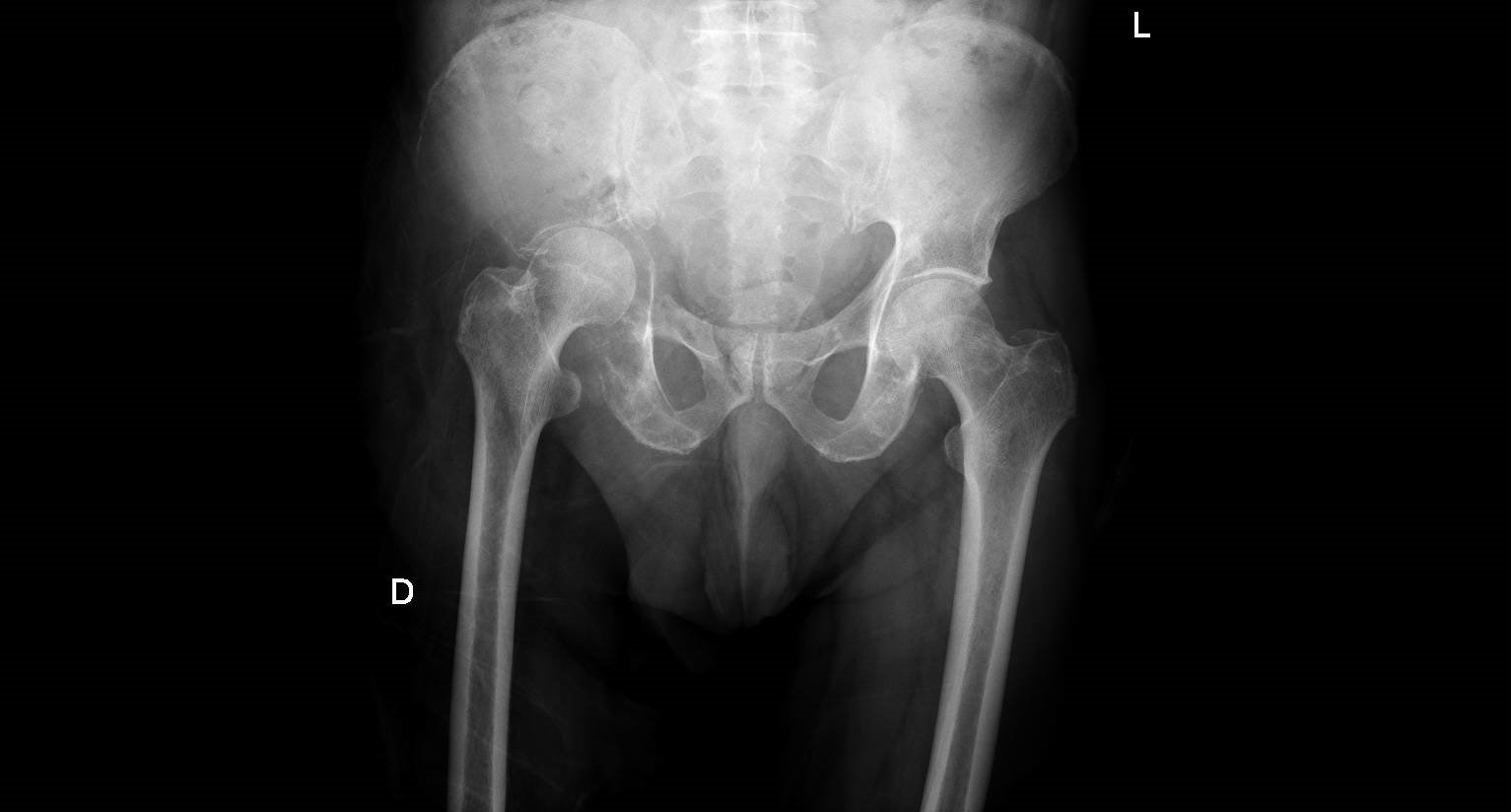
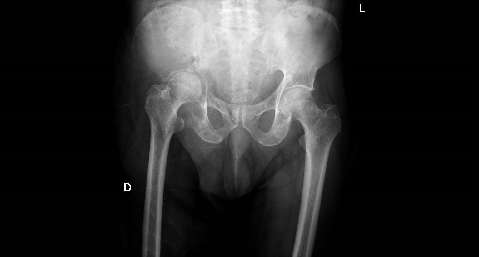
The patient was admitted with a diagnosis of respiratory infection and possible recurrence of lung cancer. No other comments were made about the patient’s pain despite the existence of a hip X-ray (no report) and which we show below

What does this image suggest? If you want to know the result, click here
Clinical evolution
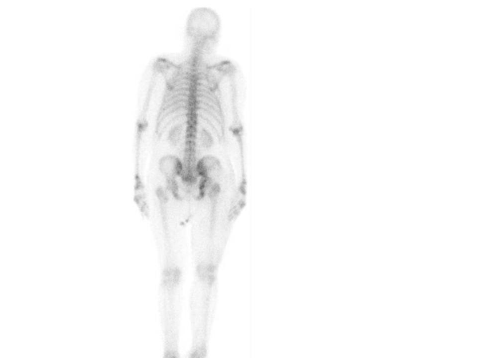
The doctor in charge of the patient considered that a new test should be requested to increase the probability of his diagnostic assumption. Of course he thought about doing an MRI, but it couldn’t be done quickly. The doctor decided to order a bone scan to find out the extent of the problem and the image is shown below

The bone scan shows increased uptake in the right hip and sacrococcygeal joint on the same side (the projection shows the right side of the body to the right of the image). If you want to see a comment and final diagnosis, click here
Lorenzo Alonso
FORO OSLER


