(Some) Radiographic patterns in Oncology and COVID-19
COVID-19 shares radiographic findings with others pulmonary diseases
Different types of lung injury, infectious and not infectious, can share similar or the same radiographic findings in different combinations. Ground-glass opacities, “crazy-paving” pattern, interlobular septal thickening and areas of consolidation (1),have been described in the CT in the COVID-19 disease, but several other diseases also share some of these findings. Moreover, a plain X-ray in COVID-19 shows an interstitial pattern, alveolar consolidation, images that can be present in other diseases. The diagnosis of COVID-19, so far, is based on the history of exposure, the clinical picture and the RT-PCR test, but even this is not 100% positive even in the presence of the disease, and the clinician must decide to test again. The real problem, dealing with this not uncommon situation in a ward with cancer patients, is to decide the attitude with the patient when you meet an image that could be associated with COVID-19, but also with the tumor or complications of the treatment. Clinical reasoning and common sense are still important
Practical approach. Clinical Case
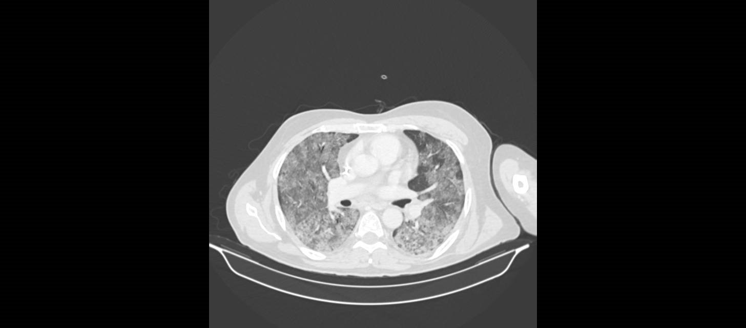
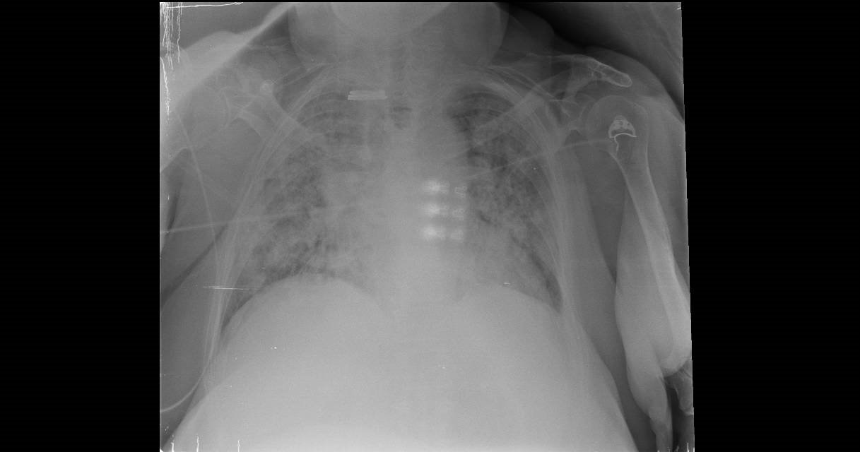
A 80-year-old man with a diagnosis of Non-Hodgkin Lymphoma diagnosed four years before. He was now in follow-up after his last cycle of chemotherapy three months ago.He was for a month with a progressive decrease in the steroid dose. Two weeks ago he started with shortness of breath, without thoracic pain, sputum or fever.
Physical examination showed hypoxemia (O2 Sat. 88% without oxygen) and mild rales in both lungs. The blood test showed a four times increase in the level of Lactic Dehydrogenase (LDH), with normal hematocrit and kidney function. A bronchoscopy was not possible due to the clinical situation of the patient.
We show here the Chest X-ray and the CT images
What would you have decided?
We started treatment with trimethoprim and sulfamethoxazole and steroids, thinking in a high probability for an infection with Pneumocistis jirovecii, based on previous steroid, the hypoxemia, an increase in LDH and the CT image.
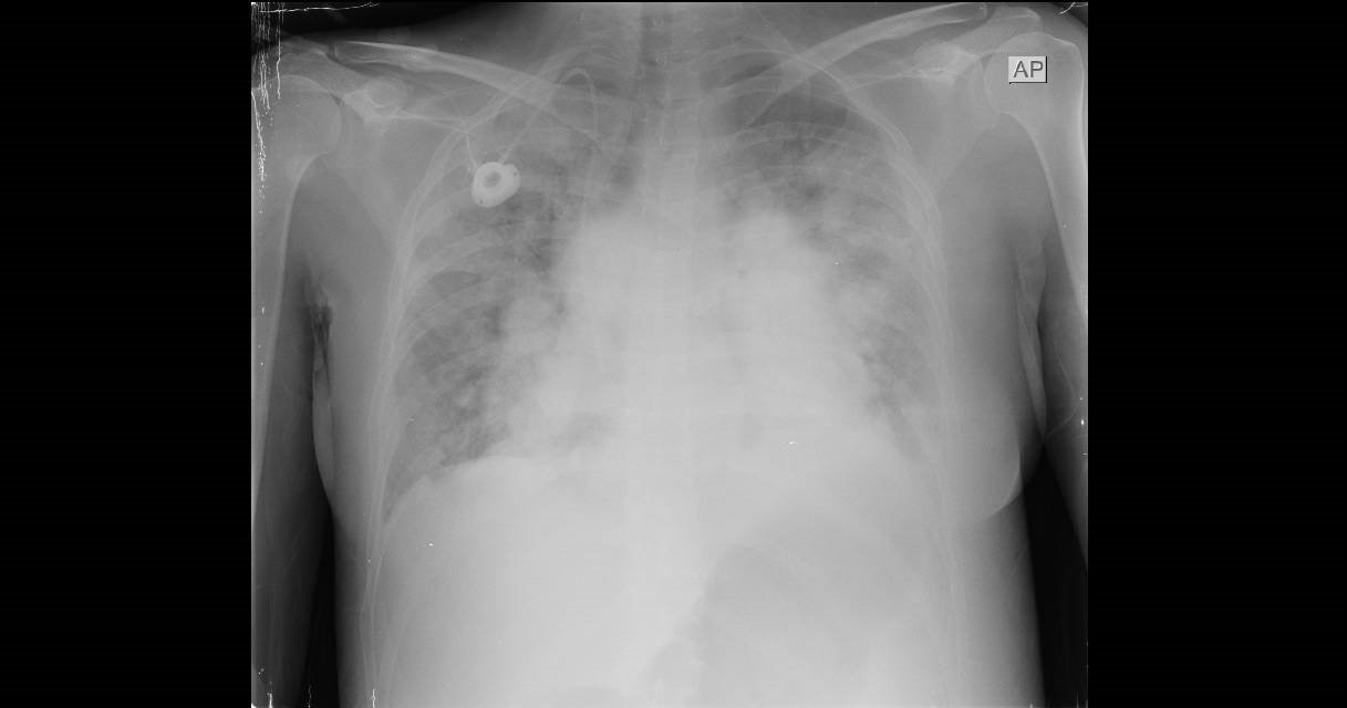
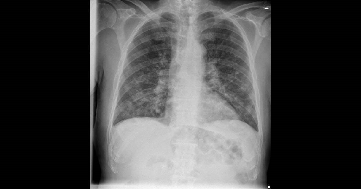
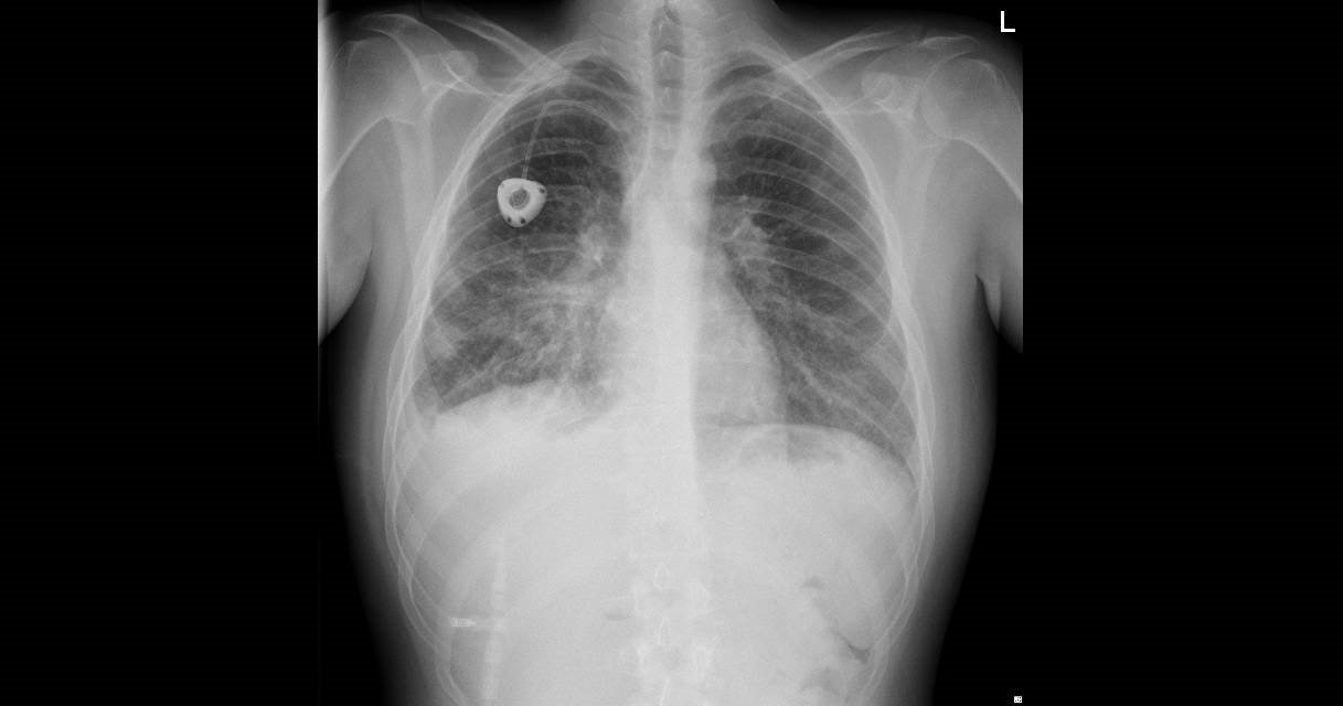
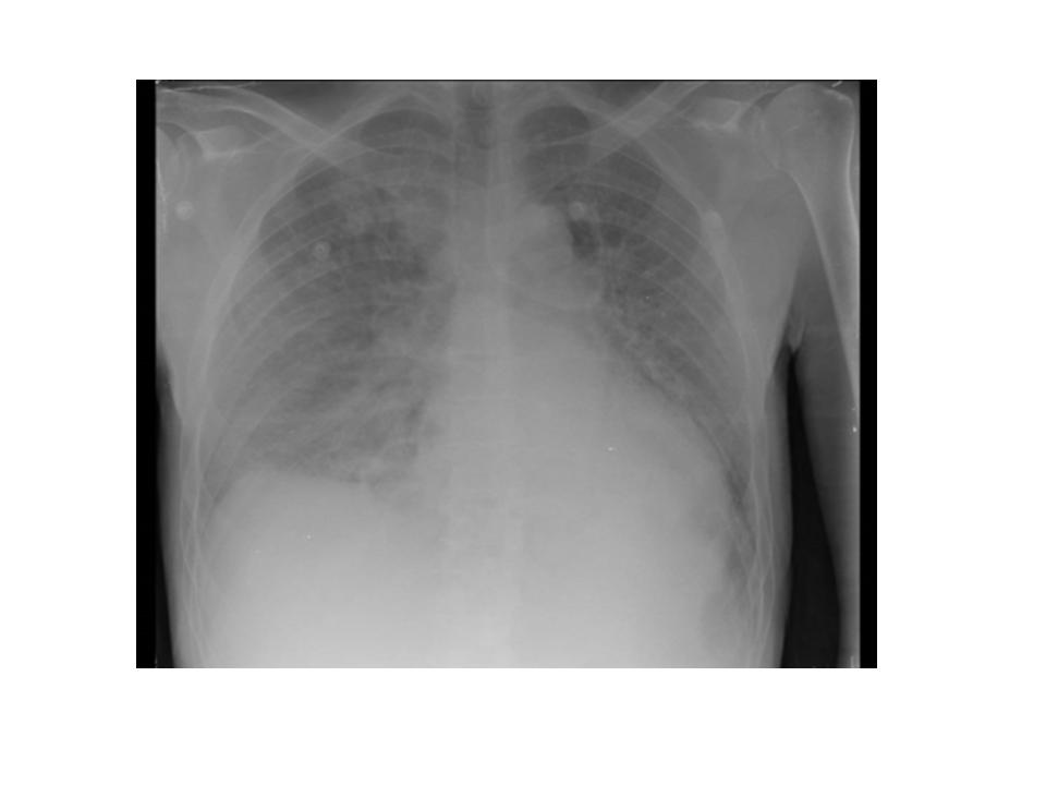
Some examples of non-COVID-19 Chest X-ray
We will show here some chest X-ray of different entities that could be included in the initial differential diagnosis of a suspected patient with the COVID-19 disease. These images are exposed here as an exercise of clinical reasoning in patients with cancer and other problems, to help to remember common situations in this group of patients.
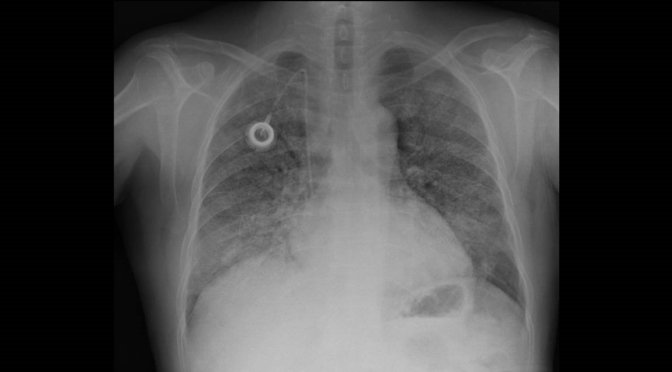
Case 1. Lymphangitis and metastases in Osteosarcoma
Case 2. Gemcitabine toxicity
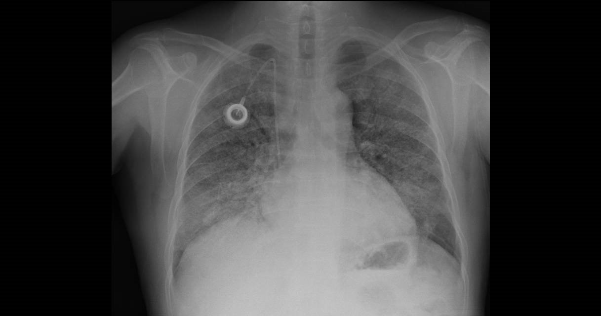
Case 3. Lymphangitis and metastases in Sarcoma
Case 4. Lenalidomide toxicity in myeloma
Case 5. Systemic sclerosis and hypertension: A) Cardiac failure and pulmonary edema B) the same patient after treatment for cardiac failure
A)
B)

Bibliography
(1) Dai WC, Zhang HW, Yu J, Xu HJ, Chen H, Luo SP, Zhang H, Liang LH, Wu XL, Lei Y, Lin F. CT Imaging and Differential Diagnosis of COVID-19. Can Assoc Radiol J. 2020 Mar 4:846537120913033. doi: 10.1177/0846537120913033. [Epub ahead of print
Author: Lorenzo Alonso, MD
FORO OSLER