A picture is worth a thousand words, more if it is definitive
A picture is worth a thousand words
Advice: all images have the patient’s consent
Looking for a sign
After the anamnesis and physical examination a doctor tries to figure out in his or her mind what is the problem with the patient. But, when time starts to run without a diagnosis, the patient, thefamily and the doctor feel uncomfortable, because the decision making process and the treatment are delayed with an increase in the possibility of a bad outcome.
Then, when a physician finds a sign that clarify the situation everyone is relief and the specific treatment can be undertaken.
We will show here two examples of this situation.
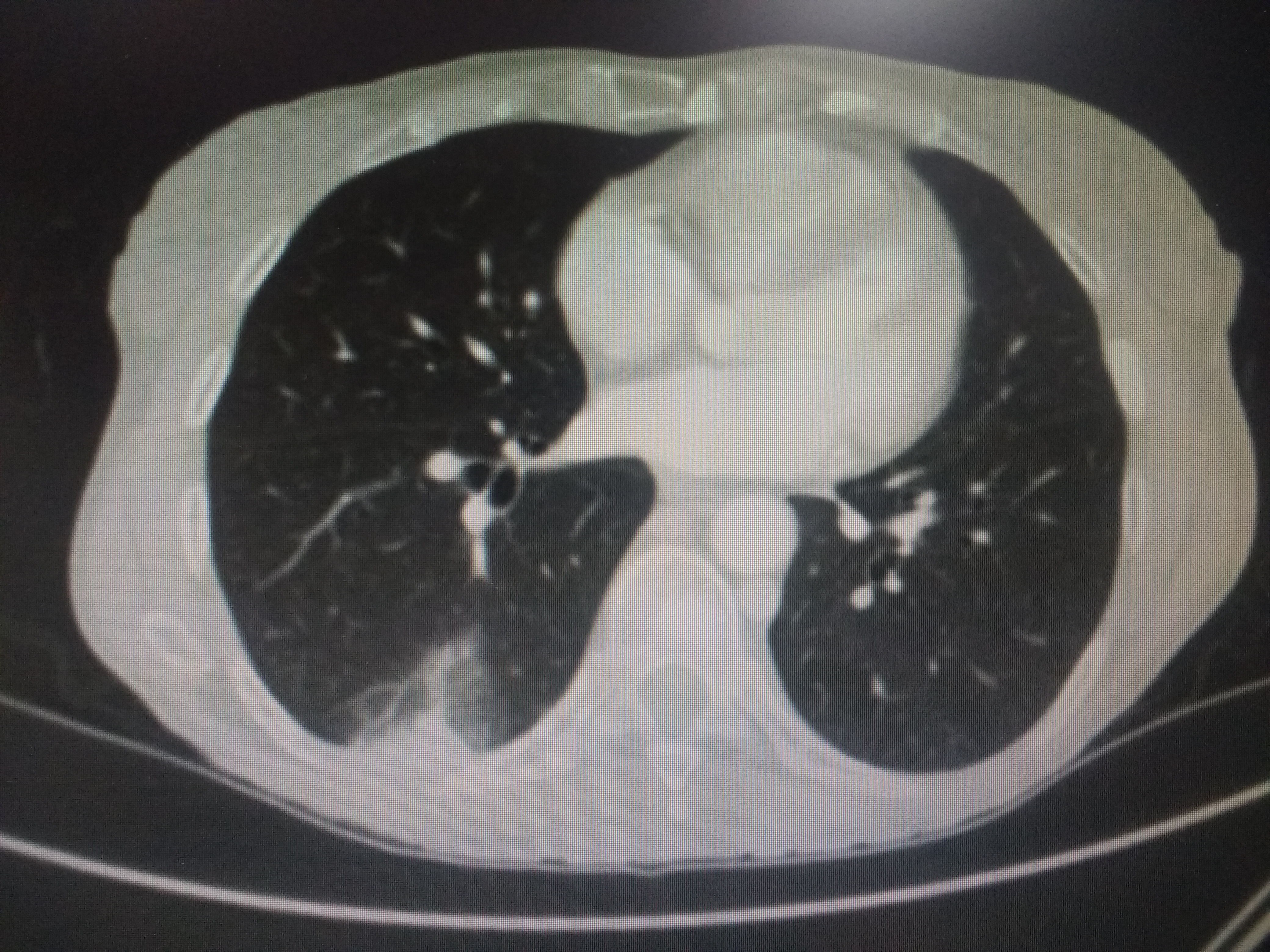
Dyspnea, thoracic pain, cough, no fever
A man with a diagnosis of colon cancer started with dyspnea while resting, cough, but no fever. Blood and sputum cultures were negative. The X-ray was not significative, but there was not a consolidation or cardiac enlargement. Basal oxygen saturation was 82%. Hemoglobin level was also normal.
Syndrome: Respiratory Insufficiency
Clinical possibilities:
- Opportunistic infection (interstitial)
- Lymphangitis
- Drug toxicity
- Pulmonary embolism
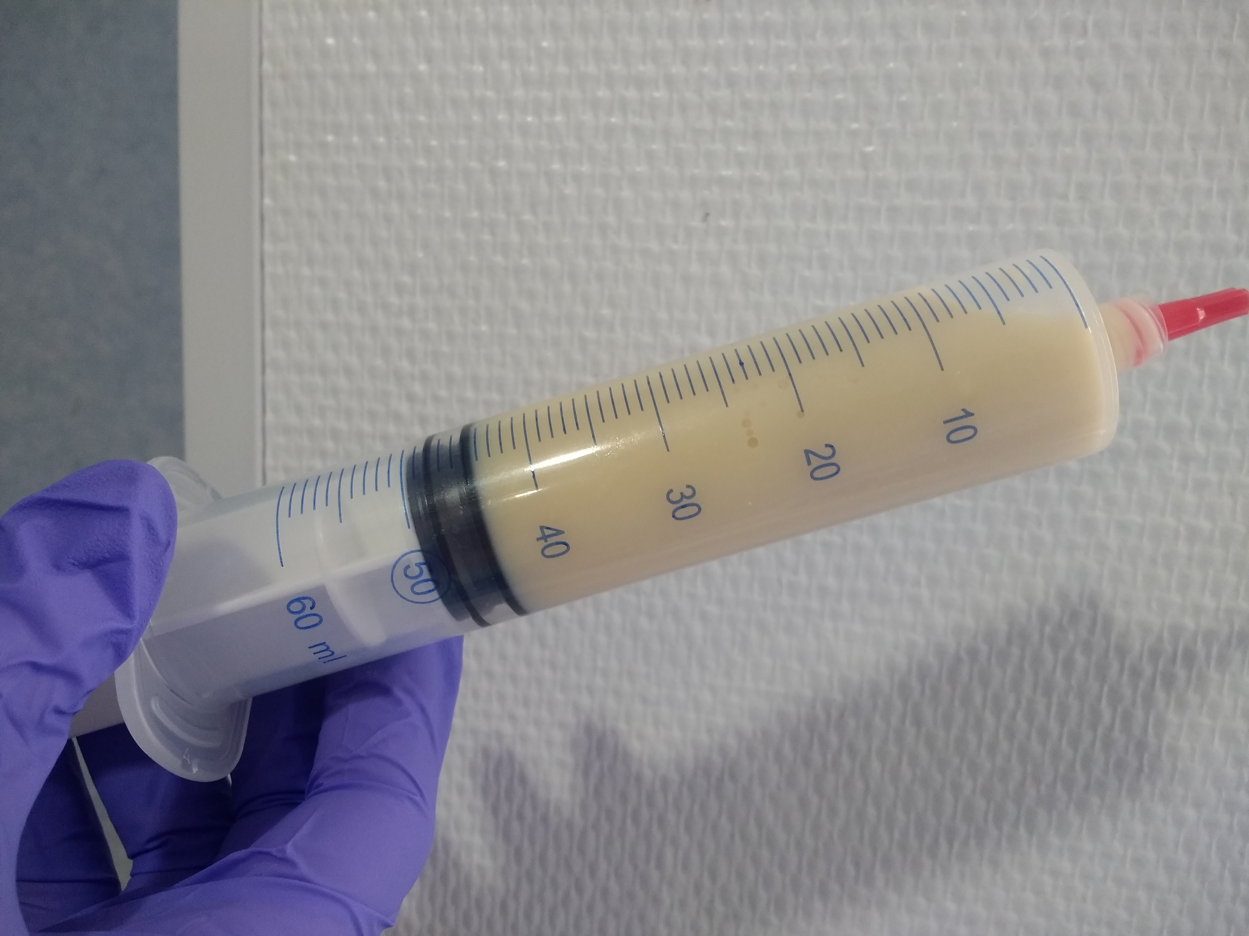
Non-Hodgkin lymphoma, ascites, no abdominal pain, no fever
A 70-year-old man had a previous diagnosis of indolent non Hodgkin lymphoma and he was under follow-úp without treatment. He had not a liver disease history and he was only taking drugs for high blood pressure.
During three months the patient started with an increase in the size of his abdomen, without pain or nausea, vomiting , constipation or fever.Liver function tests and viral markers were normal.
The sonogram was normal, including the suprahepatic veins. The CT scan showed some retroperitoneal nodes less than 1 cm in size. The liver was normal and it was clear the presence of ascites.
Syndrome: Ascites
Clinical possibilities:
- Liver disease(unknown)
- Peritoneal infection
- Lymphoma
- Venous thrombosis
ANSWER NEXT
Case: Pulmonary embolus and Pulmonary infarction with the “typical” triangular shape
Second case: Chylous ascites in relation to lymphoma (abdominal progression)